| CATEGORII DOCUMENTE |
| Alimentatie nutritie | Asistenta sociala | Cosmetica frumusete | Logopedie | Retete culinare | Sport |
PROTRUZIE ACETABULARA CU COXARTROZA BILATERALA - PREZENTARE DE CAZ
PRIMARY TOTAL HIP ARTHROPLASTY IN BILATERAL ACETABULAR PROTRUSION (Abstract): Acetabular protrusion is a clinical entity consisting of deepening of the acetabulum and consequent sinking of the femoral head within this cavity, uni- or bilaterally, such that the bottom of the acetabulum prominates in the pelvic cavity. Etiologically, the disease may be primary or secondary. This article presents the case of a 66-year old man with primary bilateral acetabular protrusion who was admitted in our clinic for a symptomatology invloving the hips: pain which was exacerbated at big efforts in orthostatic position and a progressive diminution of the amplitude of movement in both hips. The plain radiograph of the pelvis revealed the deepening of the acetabulum on both sides, intrapelvic protrusion of the bottom of the acetabular cavities, sinking of the femoral heads (and to a certain amount, also the femoral necks) into the acetabula. The articular space was altered bilaterally by osteoarthritic modifications. The diagnostic was bilateral acetabular protrusion with secondary hip osteoarthritis. Subsequently, he underwent bilateral total hip arthroplasty with an additional reinforcement with a Burch-Schneider ring on the left acetabulum. The operations were performed at a 6-months interval, with a favorable postoperative evolution for both hips. Key words: TOTAL HIP ARTHROPLASTY, ACETABULAR PROTRUSIO
Introducere
Protruzia acetabulara este o entitate clinica ce consta in infundarea capului femural la nivelul cavitatii cotiloide, uni- sau bilateral, astfel incat fundul cavitatii cotiloide devine proeminent in interiorul micului bazin. Din punct de vedere etiologic, afectiunea poate fi primara sau secundara. Forma primara, numita si arthrokatadysis sau Otto pelvis, afecteaza mai frecvent sexul feminin, se manifesta la virste nu prea inaintate si implica manifestari la nivelul ambelor solduri. Aceasta forma este un diagnostic de excludere, trebuind eliminate cele 3 posibile tipuri de etiologii: 1) afectiuni inflamatorii sau infectioase ale soldului, 2) deficite calitative ale structurii osoase acetabulare si 3) tulburari de crestere si dezvoltare cu rasunet asupra articulatiei coxofemurale. Forma secundara este rezultatul unei patologii de baza. Posibile etiologii ale protruziei acetabulare secundare sunt (1):
Infectii bacteriene (gonococice, streptococice, stafilococice, sifilis, tuberculoza) sau parazitare (echinococoza)
Afectiuni inflamatorii (artrita reumatoida, spondilartropatii, condroliza idiopatica)
Afectiuni metabolice (boala Paget, osteomalacie, hiperparatiroidism)
Afectiuni genetice (osteogeneza imperfecta, acrodisostoza, sindromul Marfan (2), sindromul Ehler-Danlos, trisomia 18, sindromul Stickler, neurofibromatoza, siclemia, homocistinuria)
Afectiuni neoplazice (primare - hemangiom, secundare - metastaze sau osteonecroze radioinduse)
Traumatisme - fracturi acetabulare
Esecul unei artroplastii totale de sold, primare sau de revizie (3); se pot folosi inele de ranforsare (4) sau cupe speciale antiprotruzie (5) in aceste operatii, in scopul prevenirii protruziei acetabulare, la pacientii cu alti factori de risc prezenti (de ex. artrita reumatoida).
Prezentarea cazului
Pacientul A.T., de sex masculin, in varsta de 66 de ani, de ocupatie agricultor, s-a prezentat in clinica noastra acuzand dureri si mai ales limitarea mobilitatii ambelor solduri, aparute in urma cu aproximativ 18 luni. Istoricul personal si familial al pacientului nu releva nici o suferinta articulara sau de natura reumatologica. In ceea ce priveste simptomatologia, durerile erau mai intense pe partea dreapta si aveau caracter mecanic, aparand la eforturi relativ mici. Limitarea mobilitatii soldurilor era de asemeni de natura mecanica, toate miscarile din aceste articulatii fiind blocate brusc peste o anumita amplitudine. De exemplu, abductia era dureroasa si brusc limitata la aproximativ 25s, datorita unui obstacol de natura osoasa (impingement). Mobilizarea pasiva intre aceste limite era insa nedureroasa.
Examenul radiologic efectuat pe radiografia de fata a bazinului (fig. 1) releva prezenta semnelor radiologice de protruzie acetabulara. Unghiul Wiberg de acoperire a capului femural (W) a avut valori de 73 pe dreapta si 84 pe stanga. Acest unghi este format intre 2 linii: o verticala prin centrul capului femural si o linie ce uneste centrul capului de spranceana cotilului si are valor normale de 25-40. Marginea interna a acetabulului este situata intern de linia ilio-ischiatica a lui Kohler, la distanta de 14 mm pe dreapta si 18 mm pe stanga (pe radiografia de fata a unui pelvis normal, marginea interna a acetabulului este situata extern de linia lui Kohler). Masurand distanta de la marginea superioara a ramului ascendent al pubisului la conturul protiunii protruzionate a acetabulului, am obtinut valorile de 11 mm pe dreapta si 15 mm pe stanga. Astfel, am incadrat leziunea pe partea dreapta ca fiind de grad II sau moderata (6-15 mm), iar pe partea stanga, de grad III sau severa (peste 15 mm).
De asemeni, se observa pensarea spatiului articular bilateral, prezenta osteofitelor si modificarile de structura ale capului femural bilateral, fapt ce denota aparitia de modificari degenerative (coxartroza bilaterala).
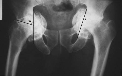
Fig. 1. Radiografia de fata a bazinului. Pe soldul drept este figurat
unghiul Wiberg (W), iar pe cel stang linia ilio-ischiatica Kohler (K).
Dat fiind caracterul bilateral, simultan evolutiv si relativ simetric al afectiunii, precum si faptului ca in urma anamnezei si a examenului clinic si paraclinic riguros nu s-a putut evidentia niciuna dintre cauzele protruziei acetabulare secundare enumerate mai sus, s-a stabilit diagnosticul de protruzie acetabulara cu coxartroza secundara bilaterala.
Optiunile terapeutice in protruzia acetabulara sunt urmatoarele: artrodeza, excizie-artroplastie, artroplastie cu proteza totala, acetabuloplastie, osteotomie intertrohanteriana de valgizare. In acest caz, luand in considerare o serie de criterii cum ar fi: maturitatea scheletului pacientului, gradul protruziei si mai ales aparitia modificarilor artrozice, am optat pentru artroplastia de sold cu proteza totala bilaterala.
Artroplastiile s-au realizat in doua interventii operatorii la distanta de 6 luni, initial pe partea dreapta (fig. 2), unde s-a instalat initial decompensarea algica si apoi pe partea stanga (fig. 3), unde a fost necesara crearea unui nou fund al acetabulului cu ajutorul unui inel de ranforsare insurubat tip Brch-Schneider. Intre inel si acetabul s-a introdus o grefa osoasa cortico-spongioasa recoltata din creasta iliaca. Pe ambele parti, componenta femurala a fost de tip necimentat.
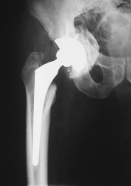
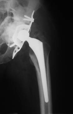
Fig. 2. Rezultatele imediate postoperatorii.
Fig. 3. Rezultat final la 1 an postoperator.
Discutii
La ora actuala, cele doua metode de tratament larg folosite in protruzia acetabulara sunt osteotomia intertrohanteriana de valgizare si artroplastia totala de sold. Osteotomia de valgizare descrisa de Pauwels (6) are ca rezultat verticalizarea directiei rezultantei fortelor ce actioneaza asupra peretelui acetabular, reducand presiunea asupra fundului acestuia si schimband totodata si zona portanta a capului femural. Rosemeyer (7) raporteaza rezultate foarte bune ale acestei metode cand este aplicata in cazuri atent selectionate (pacienti tineri, cu modificari artrozice minime sau absente). McBride (8) raporteaza de asemeni rezultate foarte bune obtinute prin aceasta metoda, dar recomanda ca procedura sa nu fie aplicata pacientilor peste 40 ani sau la cei cu leziuni artrozice evidente pe radiografie. In ceea ce priveste cazurile cu modificari degenerative in articulatie, majoritatea autorilor opteaza pentru artroplastia cu proteza totala. Sotelo-Garza si Charnley (9) nu au gasit diferente din punct de vedere al rezultatului postoperator intre soldurile protezate cu si fara protruzie acetabulara. Autorii insista asupra importantei construirii unei neoarticulatii cu centrul cat mai aproape de centrul cotilului original. Ranawat (10) a gasit deteriorarea implantului protetic la 16 din 17 cazuri in care centrul neo-articulatiei era la mai mult de 1 cm distanta de centrul articulatiei originale. La aceasta concluzie au ajuns independent si Gates (11) si Bayley (12).
In ceea ce priveste umplerea defectului dintre componenta acetabulara si peretele cotilului, MacCollum (13, 14) considera eficienta folosirea unei grefe osoase morselate, aceasta conducand si la asezarea laterala cupei intr-o pozitie cat mai apropiata de cea anatomica si de asemeni marind capitalul osos al peretelui cotiloidian. Acest procedeu este adoptat si de Rosenberg (15) pe 31 pacienti cu protruzie acetabulara secundara artritei reumatoide. Ranawat si Zahn (16), intr-un studiu pe 27 cazuri, postuleaza utilizarea doar a grefei osoase in cazurile cu protruzie peste 5 mm cu deficit osos minim al peretelui cotiloidian medial si adaugarea unui dispozitiv de fixare (ex. inel) cand acest deficit este important. In cazurile cu protruzie sub 5 mm, acesti autori nu au folosit grefa osoasa.
Bibliografie
1. Van De Velde S, Fillman R, Yandow S. The aetiology of protrusio acetabuli. Literature review from 1824 to 2006. Acta Orthop Belg 2006 Oct; 72(5):524-9.
2. Do T, Giampietro PF, Burke SW, Davis JG, Raggio C, Schneider R, Boachie-Adjei O, Brill P. The incidence of protrusio acetabuli in Marfan's syndrome and its relationship to bone mineral density. J Pediatr Orthop 2000 Nov-Dec; 20(6):718-21.
3. Hansen E, Ries MD. Revision total hip arthroplasty for large medial (protrusio) defects with a rim-fit cementless acetabular component. J Arthroplasty 2006 Jan; 21(1):72-9.
4. Gill TJ, Sledge JB, Mller ME. The Brch-Schneider anti-protrusio cage in revision total hip arthroplasty: indications, principles and long-term results. J Bone Joint Surg Br 1998 Nov; 80(6):946-53.
5. Benjamin J, Thomas M, Szivek J. The ability of various acetabular components to resist protrusio migration. Orthopedics 1997 Apr; 20(4):307-10.
6. Pauwels F, Furlong RJ, Maquet P. Biomechanics of the Normal and Diseased Hip: Theoretical Foundation, Technique and Results of Treatment - An Atlas. Berlin: Springer-Verlag, 1976, 129-169.
7. Rosemeyer B, Viernstein K, Schumann HJ. Mittelfristige Ergebnisse der Valgisierenden und Medialisierenden Intertrochanteren Osteotomie mit Verkurzung des Coxalen Femurendes bei der primaren Protrusio Acetabuli. Arch Orthop Unfall-Chir 1973;77:138-48.
8. McBride MT, Muldoon MP, Santore RF, Trousdale RT, Wenger DR. Protrusio acetabuli: diagnosis and treatment. J Am Acad Orthop Surg 2001; 9:79-88.
9. Sotelo-Garza A, Charnley J. The results of Charnley arthroplasty of the hip performed for protrusio acetabuli. Clin Orthop 1978; 132:12-18.
10. Ranawat CS, Dorr LD, Inglis AE. Total hip arthroplasty in protrusio acetabuli of rheumatoid arthritis. J Bone Joint Surg Am 1980; 62:1059-65.
11. Gates HS, Poletti SC, Callaghan JJ, McCollum DE. Radiographic measurements in protrusio acetabuli. J Arthroplasty 1989; 4:347-51.
12. Bayley JC, Christie MJ, Ewald FC, Kelley K. Long-term results of total hip arthroplasty in protrusio acetabuli. J Arthroplasty 1987; 2:275-9.
13. McCollum DE, Nunley JA. Bone grafting in acetabular protrusio: a biologic buttress. The Hip 1978; 6:124-46.
14. McCollum DE, Nunley JA, Harrelson JM. Bone-grafting in total hip replacement for acetabular protrusion. J Bone Joint Surg Am 1980; 62:1065-73.
15. Rosenberg WW, Schreurs BW, de Waal Malefijt MC, Veth RP, Slooff TJ. Impacted morsellized bone grafting and cemented primary total hip arthroplasty for acetabular protrusion in patients with rheumatoid arthritis: an 8- to 18-year follow-up study of 36 hips. Acta Orthop Scand 2000 Apr; 71(2):143-6.
16. Ranawat CS, Zahn MG. Role of bone grafting in correction of protrusio acetabuli by total hip arthroplasty. J Arthroplasty 1986; 1:131-7.
|
Politica de confidentialitate | Termeni si conditii de utilizare |

Vizualizari: 1782
Importanta: ![]()
Termeni si conditii de utilizare | Contact
© SCRIGROUP 2025 . All rights reserved